When AI Leaves the Lab: A Portable Eye Scanner Built for Community Screening
A portable scanning slit-light system shows what becomes possible when optical imaging, geometry-aware measurement, and lightweight on-device AI are designed as one complete anterior-segment screening platform.
Medical AI has a deployment gap
Artificial intelligence has become highly capable at analyzing medical images. Yet many systems remain tied to research laboratories, specialist hospitals, cloud platforms, or high-performance workstations. Strong model performance alone does not guarantee that a complete screening workflow can operate close to the people who may benefit from it.
This gap is especially relevant in eye care. Screening can help identify people who may need specialist assessment, but many established imaging systems are stationary and rely on trained personnel and centralized clinical infrastructure. People with limited access to ophthalmic services may therefore reach specialist assessment later than is desirable.
It is also “Can the entire acquisition-and-analysis system work where screening is needed?”
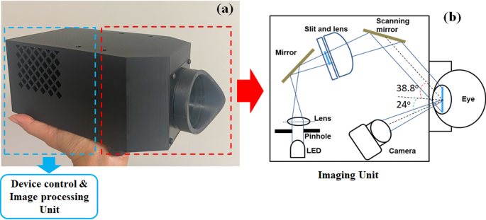
A study published in Scientific Reports examined this question through a portable, AI-integrated scanning slit-light device for imaging the anterior segment of the eye. The prototype combines motorized slit scanning, synchronized video acquisition, automated image analysis, geometric correction, and the lightweight LWBNA-unet segmentation model.
A compact platform designed around the complete workflow
The device projects a narrow slit of visible light across the front of the eye while a camera records a sequence of frames. As the slit moves, reflections from the cornea, iris, pupil region, and lens reveal structural information that can support both visualization and quantitative analysis.
The camera remains fixed while the illumination position changes. This controlled geometry matters because the software does more than classify an image: it identifies anatomical interfaces, selects useful frames, applies geometric and anatomical scaling, and estimates measurements from the segmented structures.
The study positions the prototype as a screening-oriented research platform rather than a replacement for AS-OCT, gonioscopy, pachymetry, or a complete ophthalmic examination.
How a short scanning sequence becomes quantitative information
A complete scan produces a sequence rather than a single photograph. The pipeline uses that sequence to find frames in which specific anatomical structures are most suitable for each downstream calculation.
Scan
A motorized mirror sweeps slit illumination across the anterior eye while frames are acquired.
Select
Frames are retained only when the anatomical structures required for analysis are successfully segmented.
Segment
LWBNA-unet identifies corneal and iris reflections, the pupil boundary, and corneal surfaces.
Correct
Geometry-aware calculations and per-eye scaling convert image-space distances into anatomical estimates.
Measure
Selected frames support ACD estimation and exploratory analysis of additional anterior-segment features.
Segmentation also acts as a quality-control mechanism. Frames affected by blinks, motion, defocus, or poor visibility may be rejected when the required structures cannot be delineated. Candidate frames are then ranked for measurement-specific tasks, such as identifying an iris-centered frame for anterior chamber depth or a cornea-centered frame for corneal analysis.
Why lightweight AI is part of the device architecture
Local inference
Image analysis can run on the edge device without continuous cloud connectivity.
Lower compute burden
A compact model reduces memory, processing, cooling, and power requirements.
Near-site feedback
Acquisition and analysis can occur close to the point where screening is performed.
Privacy-preserving workflow
Local processing can reduce the need to transfer identifiable imaging data to remote servers.
LWBNA-unet is a compact segmentation architecture designed to retain useful anatomical information while reducing computational demand. In this study, the model segmented corneal reflections, iris reflections, the pupil boundary, and the outer corneal surface.
The reported edge benchmark used a Jetson Orin Nano in a 15 W power configuration. A typical 51-frame scan required approximately 18.5 seconds for end-to-end processing, after an acquisition period of roughly 15 seconds. Acquisition and processing were sequential, giving a combined time of approximately 33–35 seconds per eye.
A larger network could potentially perform the same segmentation task, but increased memory, latency, cooling, or power requirements could undermine the purpose of a portable standalone system. In this setting, model efficiency is a functional engineering choice.
What the study validated—and what it did not
Approximately 170 participants were enrolled. Quantitative comparison with the CASIA-2 AS-OCT system used approximately 50 eyes with matching reference scans. The strongest validation result concerned anterior chamber depth (ACD), the study’s primary quantitative output.
Anterior chamber depth
With individualized corneal-diameter scaling, the reported Pearson correlation was r = 0.916 and Lin’s concordance correlation coefficient was 0.903. Mean bias was 0.044 mm, with 95% limits of agreement from approximately −0.300 to +0.388 mm.
Four different levels of evidence in the current study
Anterior chamber depth
ACD was directly compared with AS-OCT and showed strong correlation and concordance in the validation subset.
Central corneal thickness
CCT was estimated from the same scan, but current precision and agreement were insufficient for direct clinical substitution.
Disease-related features
- Narrow anterior-chamber anatomy
- Cataract-related lens opacity
- Corneal opacity
- Keratoconic corneal distortion
Automated disease screening
Sensitivity, specificity, external generalizability, and clinical utility for disease-level screening require larger multi-site studies.
This distinction is important. Representative cases demonstrate that the device can capture clinically recognizable features, but they do not by themselves establish diagnostic accuracy for cataract, angle-closure disease, keratoconus, or corneal opacity.
From a compact network to an integrated intelligent instrument
At Lightweight-AI, the goal is not only to reduce parameter count. The deeper objective is to design intelligence around the environment in which it must operate. In medical imaging, that means considering the full chain from controlled acquisition and image quality to quantitative analysis, hardware constraints, privacy, and referral-oriented use.
The portable scanning slit-light prototype demonstrates this philosophy in physical form. Its contribution comes from combining optical design, synchronized scanning, segmentation-based quality control, frame selection, geometric reasoning, and local inference. The AI model is one essential component of a broader measurement system.
It will also be shaped by reliable systems that can perform useful work under real-world constraints.
What must happen before routine clinical use
The current work is a promising prospective observational study, but it remains an early-stage research prototype. The sample was modest, participants were recruited from one hospital and one community screening event, and all participants were Japanese. Real-world performance may also be affected by motion, blinking, alignment, media opacity, operator technique, and environmental conditions.
Larger multi-center studies should evaluate broader demographic groups, diverse disease severity, field usability, repeatability, failure modes, referral thresholds, and disease-level screening performance. Operator training, maintenance, data governance, clinical integration, and regulatory review would also be required before routine use.
The significance of the study is therefore not that every diagnostic question has already been solved. It is that purpose-built optics and lightweight AI can be integrated into a portable system capable of standardized acquisition, automated anatomical analysis, and screening-oriented measurement.