Glaucoma: When the World Quietly Begins to Disappear
We usually think we would notice if we were losing our sight. Glaucoma is dangerous because that assumption can be wrong. It can damage the pathway between the eye and the brain quietly, while everyday vision still seems normal.
What glaucoma takes away is usually permanent. If damage continues, parts of the visual world may become less clear, seem to be missing, or become increasingly difficult to use. The purpose of screening is to detect the disease early, while useful vision can still be protected. Lightweight-AI may help identify suspicious glaucomatous changes at an earlier stage and support timely referral for specialist assessment.
Doctors can actually see part of this pathway
A fundus photograph shows the back of the eye — including the retina, retinal blood vessels, and the optic disc where nerve fibers leave the eye. In glaucoma, changes around this region may become visible before a person notices major changes in vision.
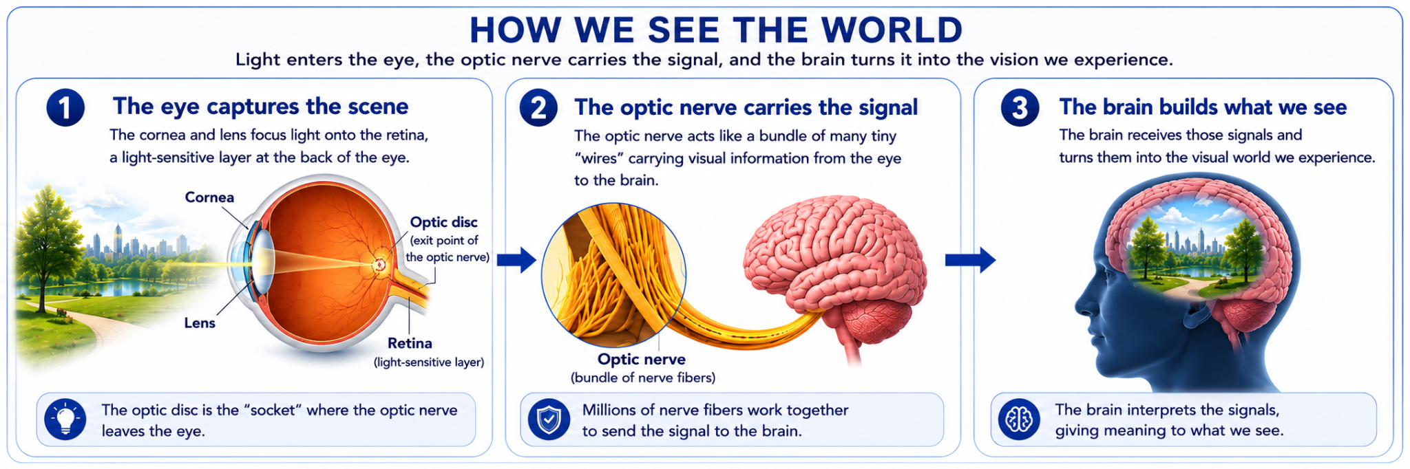
How do we see the world?
Look around you. The room, a face, a road, a tree — everything you see begins as light entering the eye.
Light is focused onto the retina, the light-sensitive layer at the back of the eye. The retina turns that light into signals.
Millions of tiny nerve fibers travel across the retina and collect at the optic disc. From there they form the optic nerve, which carries visual information toward the brain.
The brain receives these signals and interprets them as the visual world we experience.
What happens if the tiny “wires” carrying visual information to the brain are slowly damaged?
This is the frightening part of glaucoma: the world does not usually disappear all at once.
In many people, damage begins while central vision is still useful. You may still read, recognize faces, use a phone, and feel that your eyesight is normal.
Meanwhile, areas of the visual field may be becoming less clear or effectively missing, even while central vision remains useful. As more optic-nerve fibers are lost, the usable visual world becomes increasingly restricted.
Because this loss can develop gradually, the brain may adapt to missing information and the person may not immediately recognize how much of the visual field has changed.
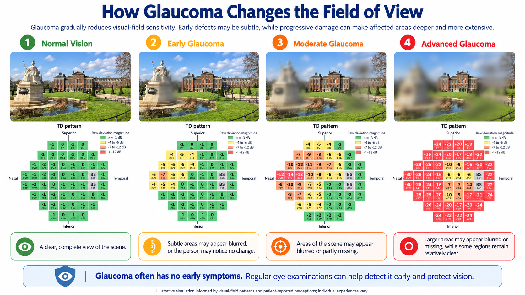
Glaucoma can slowly make parts of our visual world less clear — or seem to disappear.
Once visual-field loss has occurred because optic-nerve fibers have been permanently damaged, current treatments generally cannot restore that lost vision. Treatment aims to slow or prevent further damage. This is why detecting glaucoma before major visual loss occurs is so important.
Glaucoma can take away vision before you realize that anything is wrong.
The visual world does not usually become dark overnight. Glaucoma can damage the optic nerve quietly for years while everyday vision still feels normal. Because vision already lost is generally irreversible, the best time to detect glaucoma is before major visual loss occurs.
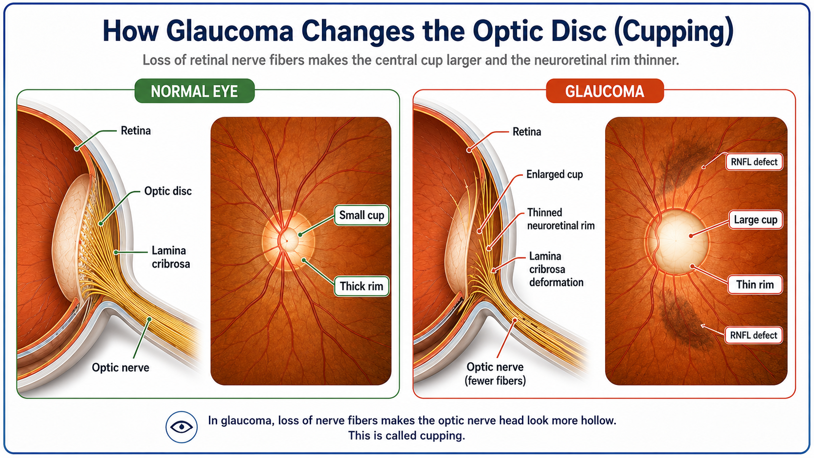
The optic nerve is slowly losing its nerve fibers
The shrinking field of vision has a physical cause: the nerve fibers carrying visual information from the retina to the brain are being damaged.
In a healthy eye, millions of retinal nerve fibers travel across the retina and converge at the optic disc, where they leave the eye and form the optic nerve.
As glaucoma damages these fibers, there is progressively less healthy nerve tissue around the optic disc.
The surrounding neuroretinal rim becomes thinner, while the central depression — the optic cup — becomes larger and deeper.
This enlargement is called optic-disc cupping.
These changes can actually be seen in a fundus photograph.
Doctors look for an enlarged optic cup, thinning of the neuroretinal rim, and areas where retinal nerve fibers have disappeared. These areas are called retinal nerve fiber layer defects (RNFLDs). Importantly, some of these structural changes may become visible before the person notices significant vision loss.
Where does eye pressure fit into this story?
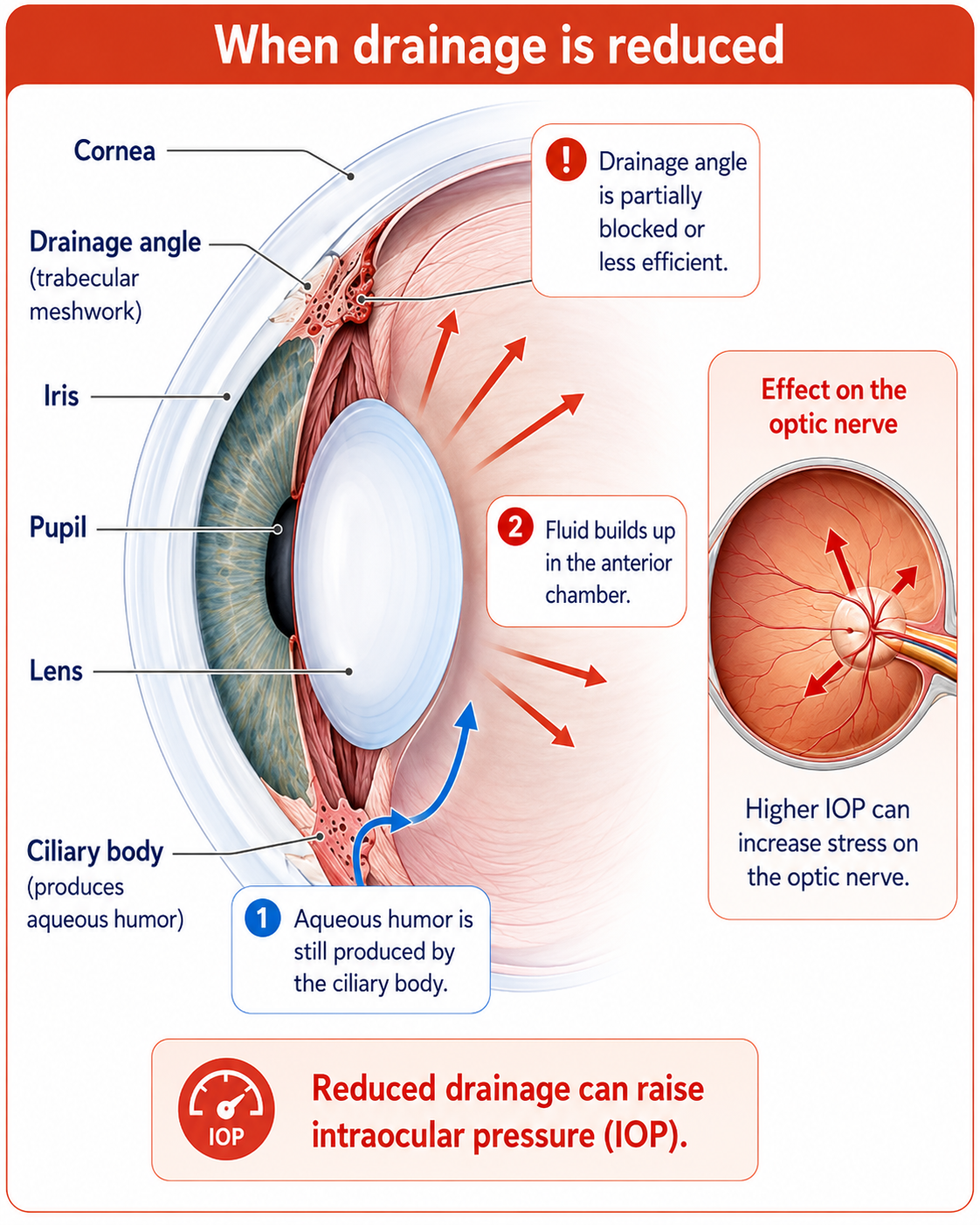
The eye is not empty. A clear fluid called aqueous humor is continuously produced inside the front of the eye and normally drains away through tiny drainage pathways.
If this fluid does not drain efficiently, it can build up and the pressure inside the eye can rise. This pressure is called intraocular pressure (IOP).
Higher eye pressure can place additional stress on the optic nerve head — the same area where millions of nerve fibers leave the eye and form the optic nerve. Over time, this can increase the risk of nerve-fiber damage and glaucoma.
But there is an important point: glaucoma is not simply “high eye pressure.” Some people develop glaucoma even when their measured eye pressure is within the usual range, while others can have elevated pressure without developing glaucomatous optic-nerve damage.
A simple way to remember it:
Eye pressure is an important risk factor. Glaucoma itself is the progressive damage occurring to the optic nerve. That is why doctors look beyond pressure and also examine the optic disc, retinal nerve fibers, and visual function.
How can glaucoma be detected before major vision is lost?
Because glaucoma can be quiet, doctors do not wait for severe symptoms. They combine several kinds of information, looking both at the structure of the eye and at how well the visual system is functioning.
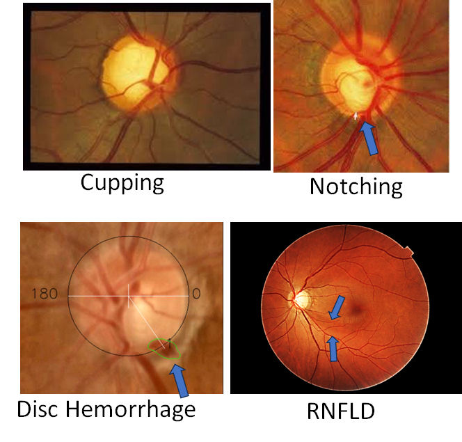
A fundus photograph can reveal warning signs of glaucoma
To most people, a fundus image may look like a simple photograph of the back of the eye. To an ophthalmologist, it contains clues about the health of the optic nerve.
Doctors examine the optic disc, the size and shape of the optic cup, and the thickness of the neuroretinal rim.
They also look for localized abnormalities such as rim notching, retinal nerve fiber layer defects (RNFLDs), and disc hemorrhage.
No single feature tells the whole story. The pattern of several findings together helps determine whether the optic nerve looks suspicious for glaucoma.
Important clues in a fundus photograph
Enlargement of the optic cup
Localized loss of neuroretinal rim
Loss of retinal nerve fibers
Small hemorrhage near the optic disc
This leads directly to AI-GS. If ophthalmologists recognize glaucoma by looking for several structural clues in a fundus photograph, then AI can be trained to look for these same clues, combine them automatically, and help identify eyes that may need closer glaucoma evaluation.
Why is early glaucoma still difficult to identify?
The fundus photograph may contain the warning signs of glaucoma, but in the earliest stages those signs can be very subtle. A small change in the optic disc, a faint RNFL defect, or a tiny disc hemorrhage can be much harder to recognize than advanced disease.
This early stage is particularly important because structural damage to the optic nerve may already be present before a standard visual-field test shows a clear abnormality. This is often referred to as pre-perimetric glaucoma (PPG).
Early changes may be difficult to judge
Advanced glaucoma may show obvious cupping and nerve-fiber loss. Early glaucoma can be very different: the abnormalities may be small, localized, or borderline.
Even experienced ophthalmologists may differ in how they interpret these subtle findings, especially when deciding whether an eye is normal, a glaucoma suspect, or showing very early disease.
A simple “glaucoma or normal?” decision can miss the details
Many conventional deep-learning screening systems treat the problem as binary classification: the entire fundus image is analyzed and the network produces a probability of glaucoma.
This can work well when disease-related changes are obvious. But when early glaucoma and normal eyes look similar, the prediction becomes more dependent on the model and the chosen decision threshold.
What does “black-box binary AI” mean?
The model may give an accurate final answer without explicitly telling us whether it recognized cupping, an RNFL defect, a disc hemorrhage, or another structural sign. More importantly, subtle early features may contribute too little to the final binary score and can therefore be overlooked.
The cases we most want to detect can also be the easiest ones to miss.
In the real-world Miyagi screening dataset used in our study, sensitivity of the standalone binary glaucoma classifier fell to approximately 56.5%. This was an important clue that simply asking one model “glaucoma or normal?” was not enough for robust screening.
This raised a different question: instead of asking one AI model only “Is this glaucoma?”, could we build a system that deliberately looks for the same individual warning signs that ophthalmologists examine — cupping, RNFL defects, disc hemorrhage and other optic-disc features — and then combines all of that evidence?
AI-GS: look for the glaucoma clues, not only the final label
The previous problem suggests a different way to use artificial intelligence. Instead of asking one model only “Is this glaucoma or normal?”, could several lightweight AI models deliberately examine the individual warning signs of glaucoma and then combine that evidence?
This was the idea behind the AI-based Glaucoma Screening network (AI-GS). Rather than relying only on one black-box probability, AI-GS combines information from several specialized lightweight models and reports clinically recognizable structural findings.
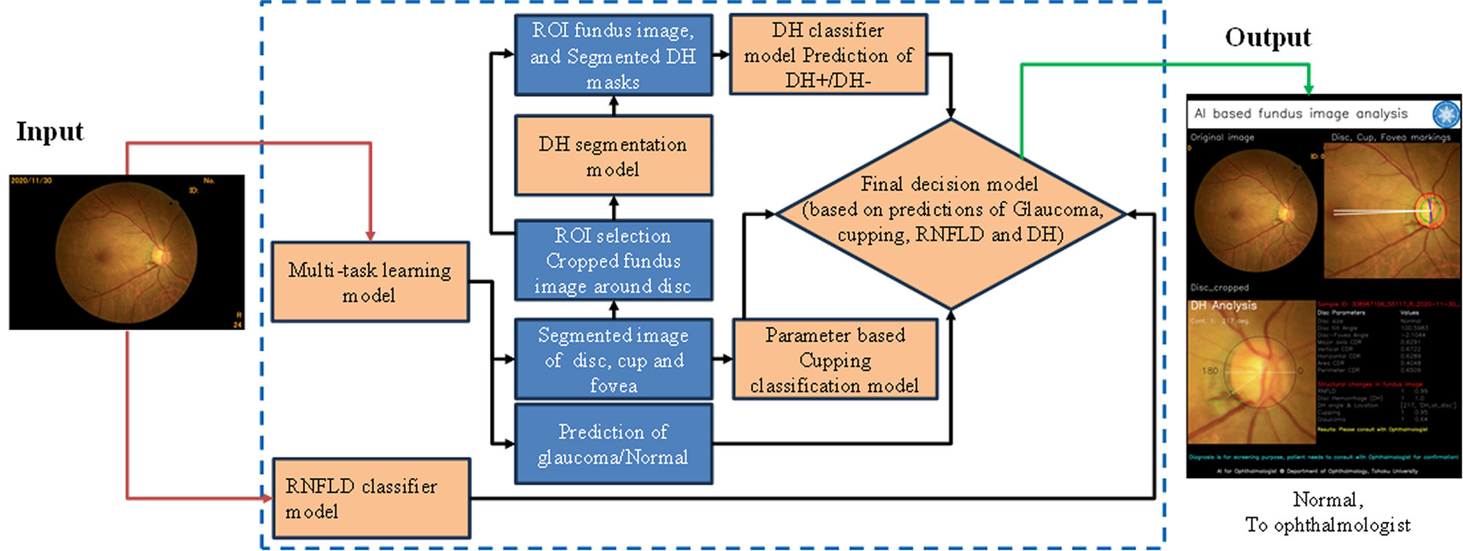
Look for subtle structural clues
Specialized models search for retinal nerve fiber layer defects (RNFLDs) and disc hemorrhage — findings that can be important when glaucoma is still subtle.
Measure the optic disc and cup
Segmentation provides cupping-related measurements and other optic-disc parameters, adding quantitative information rather than relying only on a visual impression.
Combine multiple pieces of evidence
Structural findings are combined with direct deep-learning glaucoma predictions to generate a final referral-oriented screening decision.
Tested in two very different situations
AI-GS was evaluated first on a balanced testing dataset with established glaucoma and non-glaucoma labels, and then in a much more difficult real-world screening population. These settings answer different questions, so the percentages should not be interpreted as directly equivalent tests.
Can AI-GS recognize glaucoma in a well-defined test set?
The testing dataset contained 8,370 fundus images: 4,185 glaucoma and 4,185 non-glaucoma images. None of these images had been used for model training or validation.
This experiment asks whether AI-GS can recognize established glaucoma patterns when evaluated against a balanced dataset with defined reference labels.
What happens when screening cases are less clearly defined?
The Miyagi screening data included 10,767 images marked normal and 529 glaucoma-labeled images. Glaucoma and normal labels were assigned by screening ophthalmologists from the screening fundus photographs.
This was not a complete glaucoma diagnostic work-up. The study reports that detailed diagnostic procedures such as perimetry/visual-field testing and IOP measurements were unavailable for this glaucoma group. The Miyagi labels were assigned by screening ophthalmologists from the screening fundus photographs.
In this harder setting, AI-GS increased sensitivity from 56.5% with the standalone binary classifier to 80.5%, while maintaining 91.1% specificity.
Why should 94% and 80.5% not be compared as if they were the same test?
The balanced testing dataset asks whether AI-GS can recognize glaucoma against established labels. Real-world screening asks a harder and somewhat different question: can the system identify eyes that look suspicious when the abnormalities may be subtle and the reference judgment itself is based mainly on what can be seen in the fundus photograph?
The important real-world finding: when a single binary classifier struggled in screening, combining several glaucoma-related clues substantially improved sensitivity. This supports the central idea of AI-GS: look for multiple pieces of glaucoma evidence, rather than depending on one black-box probability alone.
Do not wait for glaucoma to announce itself.
With many diseases, pain or obvious symptoms warn us that something is wrong. Glaucoma can be different. A person may still read, recognize faces and feel that everyday vision is normal while optic-nerve damage is already developing.
That is why glaucoma screening and eye assessment matter before symptoms become obvious. Eye pressure, fundus examination, OCT and visual-field testing each reveal a different part of the disease story.
A fundus photograph is especially useful for screening because it is practical and can reveal optic-disc cupping, RNFL defects and disc hemorrhage. AI-GS explores how lightweight AI can examine these clues together and help identify eyes that deserve closer specialist evaluation.
We see through millions of nerve fibers; glaucoma can silently damage those fibers; lost vision is usually permanent — so the best time to find glaucoma is before the world begins to disappear.
Where AI-GS fits: AI-GS is a research screening system, not a replacement for an ophthalmologist and not a definitive glaucoma diagnosis. The published study emphasizes that glaucoma diagnosis requires broader clinical assessment and that visual-field testing remains essential for confirming functional damage. AI-GS is designed to help identify suspicious eyes earlier and support referral for a complete examination.
Want to see how the system works? Try the online AI-GS web application or read the published npj Digital Medicine study.
Online research demonstration: AI-GS web application